Cambio - Excellence in Molecular Biology

Search
Isothermal Nucleic Acid Amplification

Isothermal Nucleic Acid Amplification: RPA Optimization Enzymes & Kits

Isothermal Nucleic Acid Amplification Kits, Enzymes, and Lysis Solutions


Since 2016, Intact Genomics has pioneered the development and manufacture of products for isothermal nucleic acid amplification. Our comprehensive portfolio includes RPA master mixes and kits, individual RPA enzymes, glycerol-free and lyophilized formats, FastAmp® Viral and Cell Lysis Solutions, and helicase-based amplification enzymes. Learn more by downloading our white paper on RPA technology and Intact Genomics RPA products.

Designed for rapid, sensitive nucleic acid amplification without conventional thermal cycling, our products support molecular diagnostic development, point-of-care and agricultural testing, and life science research. Through continued innovation, consistent quality, and reliable performance, we help researchers and industry partners accelerate discovery and develop next-generation testing solutions. Bulk quantities, custom formulations, CDMO and OEM services are also available.

Product Links

Package Size

Features

IG® RPA Master Mix

25 rxns, 100 rxns, 500 rxns

Delivers powerful isothermal amplification for molecular diagnostics and research. Easy-to-use, fast, reliable, and cost-effective.

IG® RPA Master Mix - Lyophilized(opens in new tab)

24 rxns, 96 rxns, 480 rxns

A ready-to-use, freeze-dried premix for rapid and sensitive isothermal DNA amplification.

IG® Recombinase Polymerase Amplification (RPA) Kit v2

25 rxns, 100 rxns, 500 rxns

Provides faster and more discernible results for isothermal DNA amplification.

FastAmp® qRPA SYBR Kit

25 rxns, 100 rxns, 500 rxns

Screens primer conditions to minimize non-template control (NTC) background.

FastAmp® Plant Direct RPA Kit

25 rxns, 100 rxns, 500 rxns

Rapid DNA amplification from plant tissues at a constant temperature of 37°C.

IG® Recombinase Polymerase Amplification (RPA) Kit

25 rxns, 100 rxns, 500 rxns

Contains the reagents and enzymes needed for DNA amplification.

T4 UvsX DNA Recombinase(opens in new tab)

100 μg, 500 μg, 1000 μg

Important for error-free repair of DNA double-strand breaks and replication fork restart.

T4 UvsY Protein

100 μg, 500 μg, 1000 μg

Phage T4 recombination mediator protein that stimulates filament nucleation.

T4 gp32 Protein

100 μg, 200 μg, 500 μg

Single-stranded DNA-binding protein involved in DNA replication, recombination, and repair.

Bsu DNA Polymerase Large Fragment

1,000 units

Polymerase useful for extending primers in RPA.

Sau DNA Polymerase Large Fragment

1,000 units

Polymerase useful for extending primers in RPA.

Glycerol Free or Lyophilized T4 UvsX DNA Recombinase

100 μg, 500 μg, 1000 μg

Supports error-free DNA double-strand break repair and replication fork restart.

Glycerol Free or Lyophilized T4 UvsY Protein

100 μg, 500 μg, 1000 μg

Recombination mediator protein that stimulates filament nucleation.

Glycerol Free or Lyophilized T4 gp32 Protein

500 μg

Single-stranded DNA-binding protein for DNA replication, recombination, and repair.

Glycerol Free or Lyophilized Bsu DNA Polymerase Large Fragment

1,000 units

Polymerase useful for extending primers in RPA.

Exonuclease III

10,000 units, 25,000 units

Digests one strand of duplex DNA from blunt ends, 5′ overhangs, or nicks.

Endonuclease IV (Nfo)

2,000 units, 5,000 units

Functions as an apurinic/apyrimidinic nuclease and 3′-terminal di-esterase for DNA repair.

FastAmp® Viral and Cell Lysis Solution (1×, 5×)

25 reactions, 100 reactions

Enables direct use of lysed DNA or RNA in RPA, RT-isothermal amplification, PCR, and RT-qPCR without extraction.

Tte MutL Protein(opens in new tab)

(opens in new tab)20 µg, 400 µg

A thermostable ATPase accessory protein that interacts with Tte UvrD helicase, suitable for mismatch repair studies, helicase regulation, and isothermal DNA amplification development.

Tte UvrD Helicase(opens in new tab)

2 µg, 40 µg

A thermostable, ATP-dependent DNA helicase and key enzyme for HDA, suitable for isothermal nucleic acid amplification, DNA-unwinding studies, helicase activity assays, and primer- or probe-based detection workflows.

igSuper™ Tte UvrD Helicase(opens in new tab)

(opens in new tab)2 µg, 40 µg

An engineered, thermostable ATP-dependent DNA helicase for HDA and other isothermal nucleic acid amplification, DNA-unwinding studies, and DNA detection workflows.


T4 UvsY Protein

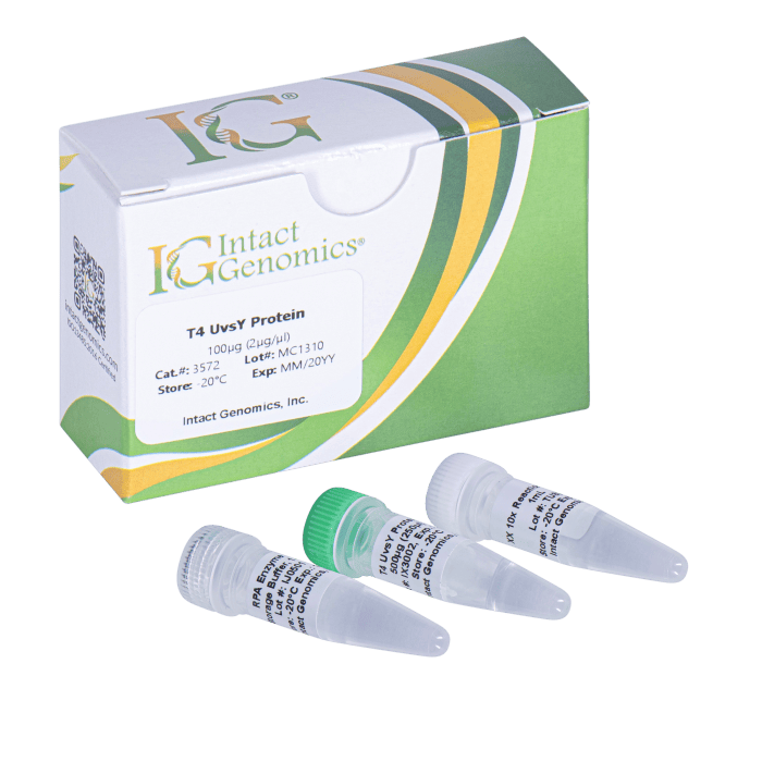
T4 UvsY Protein is a recombination mediator that enhances UvsX loading onto single-stranded DNA by displacing gp32. It stimulates UvsX ATPase activity, filament formation, strand invasion, and efficient homologous recombination.
Intact Genomics

Catalogue No.DescriptionPack SizePriceQty
  • Change to:
IG-3572T4 UvsY Protein100µg (2µg/µl) �330.82 Quantity
Add
IG-3575T4 UvsY Protein500µg (2µg/µl) �988.89 Quantity
Add
IG-3577T4 UvsY Protein1000µg (2µg/µl) �1,488.69 Quantity
Add

T4 UvsY Protein


T4 UvsY Protein is a recombination mediator that enhances UvsX loading onto single-stranded DNA by displacing gp32. It stimulates UvsX ATPase activity, filament formation, strand invasion, and efficient homologous recombination.

Product Description

UvsY is the phage T4 recombination mediator protein, and structural and biophysical studies provide insights into its role in T4 homologous recombination. During T4 homologous recombination, the UvsX recombinase must compete with the prebound gp32 single-stranded binding protein for DNA-binding sites and T4 UvsY Protein stimulates this filament nucleation event (1). UvsY is a 15.8-kDa protein with properties that are consistent with its role as mediator; it stimulates the DNA-dependent ATPase activity of UvsX, lowers the critical concentration of UvsX that is required for activity, and promotes strand exchange (2, 3). UvsY efficiently promotes the UvsX-catalyzed strand invasion reaction by recruiting to ssDNA–gp32 complexes, promotes the release of gp32, and favors the binding of UvsX.

Protein Purity

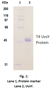
The physical purity of this enzyme is ≥98% as assessed by SDS-PAGE with Coomassie® blue staining (Fig. 1).
Product Source

E. coli BL21 (DE3) strain expressing T4 UvsY gene with an N-terminal His-tag.

This His tag does not have any effect on UvsY activity.

Product Includes

UvsY protein
10X UvsY Reaction Buffer

1x UvsY reaction buffer composition

20 mM Tris-acetate, 100 mM Potassium acetate, 10 mM Magnesium acetate, 1 mM DTT, pH 7.8 @ 25ºC

Storage Buffer

50 mM Tris-HCl, 50 mM KCl, 1 mM DTT, 0.1 mM EDTA, 50% Glycerol, pH 7.5 @ 25ºC

Storage Temperature
-20ºC

Quality Control assays
UvsY is free from detectable nuclease activities.

References

1. Beernink HT, Morrical SW. RMPs: Recombination/replication mediator proteins. Trends Biochem Sci. 1999;24(10):385–389.

2. Hashimoto K, Yonesaki T. The characterization of a complex of three bacteriophage T4 recombination proteins, uvsX protein, uvsY protein, and gene 32 protein, on single-stranded DNA. J Biol Chem. 1991;266(8):4883–4888.

3. Beernink HT, Morrical SW. The uvsY recombination protein of bacteriophage T4 forms hexamers in the presence and absence of single-stranded DNA. Biochemistry. 1998;37(16):5673–5681


Protein Concentration 


Intact Genomics Concentration Determination Method
IG uses orthogonal, 3-part approaches to determine the enzyme concentration to provide you with consistent and reliable enzymes for your needs. The quantity of a protein sample is assessed using densitometry with polyacrylamide gel electrophoresis (PAGE), UV absorbance spectra of native protein, and using a protein standard assay such as bicinchoninic acid assay (BCA) using bovine serum albumin (BSA) as a standard (Figure 1).
 

Why Does IG Use All Three Approaches?
1. Each method above has limitations. The limitations include experimental noise, accuracy, and susceptibility to buffer and/or enzyme conditions.
2. Each enzyme has unique physical properties that make a single approach to analyzing proteins a challenge. Each enzyme has a different protein sequence, different requirements to be stable in solution, and different requirements to retain its maximal activity. These differences can interfere with or convolute results, especially when compared to other enzymes. When used together, however, each method provides the scientist with independent measures of both enzyme and buffer purity and quality.
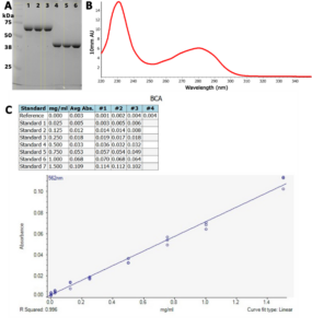
Figure 1: Enzyme quantitation methods used by IG. A) SDS-polyacrylamide gel electrophoresis. Ladder in 1st lane, 2 µg BSA (~67 kDa) as a standard in lanes 1-3, and IG enzymes (~40 kDa) in lanes 4-6. The yellow boxes are the areas evaluated by densitometry. The integrated band intensities of IG enzymes are compared with integrated band intensities from BSA to assay concentration. B) UV spectrum of a clean IG enzyme with protein peaks at 230 nm and at 280 nm. An extinction coefficient at 280 nm is typically used to quantify protein using these spectra with buffer subtraction at 330 nm. C) BCA standard curve for BSA. The curve is used to calculate an IG enzyme concentration using BSA as the standard.


Manual – T4 UvsY Protein

MSDS – IG T4 UvsY Protein


If you cannot find the answer to your problem then please contact us or telephone +44 (0)1954 210 200