Highlights
- Ideal optical properties for transmitted light and fluorescence microscopy
- Visualization of transfection process in living cells
- Simple transfection protocol (no optimization necessary)
- DNA and siRNA transfection
- Cells can be fixed and dyed
- Compatible with oil immersion
μ-Transfection Kit VI FluoR is the ideal
solution where visualization of the carrier system's path through the
cell is necessary, for example to gain information about the
transfection process.
In addition to all the benefits of the
μ-Transfection Kit VI FluoR, this kit offers the option of tracking
lipoplexes in living cells. For this purpose the transfection reagent
METAFECTENE® μ was labeled with the fluorescent dye rhodamine as
METAFECTENE® μ FluoR, while retaining its other properties. This enables
the transfection process itself to be tracked microscopically.

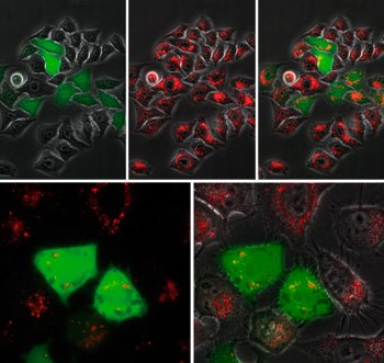
High-resolution microscopic view of HepG2 cells transfected with the
μ-Transfection Kit VI FluoR. The images clearly show the fluorescent
red-marked lipoplexes taken up by endocytosis and the incipient
expression of the fluorescent green GFP. (Images of the individual
fluorescence channels and layered images).
If you cannot find the answer to your problem then please contact us or telephone +44 (0)1954 210 200