Rapid MicroNucleus MoA –
Fast Detection of True Positive Compounds
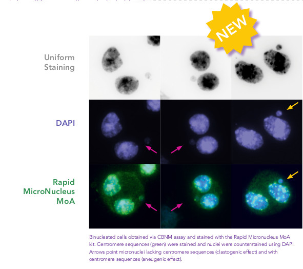
Fastest FISH assay ever! - Fast, clear identification of Aneugens and Clastogens
Introducing the new in vitro Human cell micronucleus test ( MNvit), that gives you results in 1 hour. MNvit is one of the most widely used in vitro genotoxcity assays combined with, or following the AMES test.
This is a method that provides a comprehensive basis for investigating chromosome damaging potential of a chemical or a radiation. The Patent pending MicroNucleus MoA kit provides additional information on the mechanisms of chromosome damage and micronucleus formation: aneugens and clastogens.
It is also able to provide information on Chromosomal aberrationsand Identify agents that cause structural aberrations in cultured mammalian cells. The kit uses a very simple protocol with minimal additional requirements and equipment already present in many laboratory environments.
Key features
- 1 hour FISH Assay – fast, reliable genotoxicity assessment
- High specificity – no false positive results due to RNA, low-density micronuclei, precipitates, cytoplasmic alterations
- Ready to use kit including pan-centromeric probes, DAPI and reagents
- Simultaneous DAPI Counterstain
- Fast, clear identification of mode of action: Aneugens or Clastogens
- FISH staining of centromeres is recommended in OECD TG487
- Can be used for Chemicals, Pharmaceuticals, Cosmetics, environmental samples
- For all human cells and whole blood
Overview of Procedure Rapid
Micronucleus MoA is a ready to use kit for the fast identification of
aneugens and clastogens according to OECD 487. Cells are prepared as per
standard protocols according to OECD TG 487 and the slides are
sequentially treated with a series of solutions provided with the kit
“Rapid Micronucleus MoA”, including a human-specific, highly sensitive
centromere probe and a counterstaining solution containing DAPI
(4’,6-diamidino-2-phenylindole). Slides are sequentially washed with PBS
and an ethanol series after treatment. Centromere probes are then added
to the area of interest and the slide is heated at 80°C for 3 minutes
and transferred to a humidified chamber for 20 minutes. Washes and a
5-minutes counterstaining step follow prior to the addition of the
mounting solution.
Using a 40× magnification objective it will be possible to easily
identify micronuclei and assess whether a centromere is present.
Automated reading and archiving can be obtained with the slide scanning
systems, e.g. Metafer.
Slides are usually ready for evaluation within 1 hour. The application
of FISH probes allows directly to distinguish micronuclei originating
either from chromosome loss (aneugenic compounds) or breakage
(clastogenic compounds).
FISH probe technology as used in the
Rapid Micronucleus MoA Kit can also be used in chromosomal aberration
tests (OECD 473), cytogenetic analysis in Research or in physical
dosimetry, i.e. people exposed in medical or natural radiation.
The frequency of micronuclei has been
used for many years as a biomarker to measure chromosomal damage
according to guideline OECD TG487.
References
- Cuceu C, Colicchio B, Jeandidier E, Junker S, Plassa F, Shim G,
Mika J, Frenzel M, Al Jawhari M., Hempel WM, O'Brien G, Lenain A, Morat
L, Girinsky T, Dieterlen A, Polanska J, Badie C, Carde P, M'Kacher R.
2018. Independent mechanisms lead to genomic instability in Hodgkin
lymphoma: Microsatellite or Chromosomal. Cancers (Basel).
10(7).pii:E233. Erratum in: 2019. Cancers (Basel). 11(6).pii:E757.
- Zaguia N., Laplagne E., Colicchio B., Cariou O., Al Jawhari M.,
Heidingsfelder L., Hempel WM, Bel Hadj Jrad B, Jeandidier E, Dieterlen
A, Carde P, Voisin P, M’Kacher R. 2020. A new tool for genotoxic risk
assessment: Reevaluation of the cytokinesis block micronucleus assay
using semi-automated scoring following telomere and centromere staining.
Mutat Res Gen Tox En. 850-851:503143.
If you cannot find the answer to your problem then please contact us or telephone +44 (0)1954 210 200